0

Nasopalatine duct cyst.

*Non odontogenic cysts*
1)Nasopalatine duct cyst.

-It's the commonest of nonodontogenic cysts,also known as incisive canal cyst and may be seen in the palatal soft tissue and so known as cyst of palatine papilla.
-Median palatine cyst (median cyst of the maxilla) may represent a posteriory displaced naso- palatine duct cyst.
-Nasopalatine ,incisive canal cyst ,cyst of palatine papilla and median alveolar cyst are all variants of the same lesion varying in position.

Etiology & pathogenesis,
-It is a developmental cyst arises from remnants of nasopalatine duct which connects oral and nasal cavities in the embryo, trauma or infection may be the cause that stimulate cystic degeneration.
-The cyst occurs anywhere along the duct's course which runs from the posterior palatal midline to the soft tissue of palatine papilla (incisive papilla), cyst in posterior palatine midline termed median palatine cyst and those formed in incisive papilla termed cyst of palatine papilla and all are considered nasopalatine duct cyst.


For further information about location and morphology of nasopalatine duct,follow or download this link,
http://archotol.ama-assn.org/cgi/reprint/126/6/741.pdf


Clinical features,
-Males are more affected.
-Many lesions are asymptomatic and discovered on routine radiographic examination , but may present as a swelling in soft tissue palatine papilla or may in midline of palate.
-Such lesions may discharge into the mouth through a sinus with a salty taste.


Radiographic picture,
-Well-defined round or ovoid or heart shape radiolucency at midline or may displace to one side.
-Anterior nasal spine superimposed on the lucent area giving the appearance of heart shape radiolucency.
-Diameter of nasopalatine cyst is in the range from 6mm to 6cm, so small cysts are difficult to be distinguished from incisive foramen which is of normal size from 6 to 7 mm in diameter.
-In rare instances , a nasopalatine duct cyst may develop in the soft tissues of the Incisive papilla area without anybony involvement. Such lesions often are called cysts of the incisive papilla . These cysts frequently demonstrate bluish discoloration as a result of the fluid content in the cyst lumen .


Histopathological features,
-The epithelial lining of nasopalatine duct cysts is highly variable, it may be stratified squamous epithelium or pseudostratifies ciliated columnar epithelium and less frequently simple columnar or cuboidal epithelium.
-Cyst arises in the superior aspect of the canal near nasal cavity is more likely to be respiratory epithelium (pseudostratified ciliated columnar epithelium) and cysts arising in the inferior position near the oral cavity are more likely to exhibit stratified squamous epithelium.
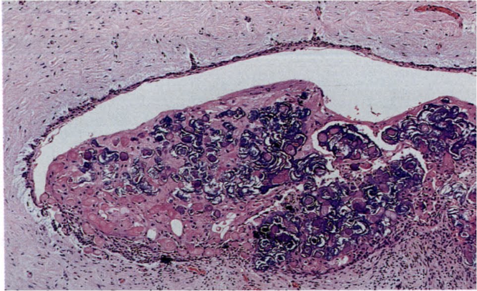
-Mucous secreting cells may be present and connective tissue wall also contains small arteries and nerve tissue,also an inflammatory response may be noted in the cyst wall

Simple cuboidal lining
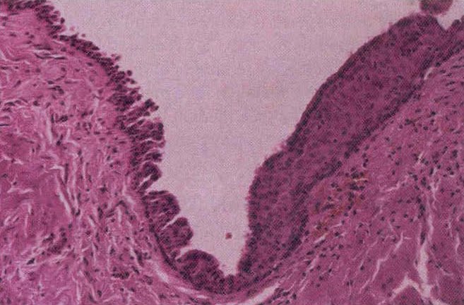
Pseudo stratified columnar and stratified squamous

Show inflammation

Arteries & nerves

Treatment,
Surgical enucleation without recurrence.

تنزيل كتابCawson's essentials of oral pathology and oral medicine


R.A.Cawson.Cawson’s essential Oral pathology&oral medicine,7th edition,Churchill livingstone,Spain;2002

حمل من هنا
Choose your link

(91.36 MB)


http://www.multiupload.com/MU_KOIY2CY7E7http://www.multiupload.com/DF_KOIY2CY7E7

http://www.multiupload.com/ZS_KOIY2CY7E7

OR

http://www.multiupload.com/KOIY2CY7E7

0

تنزيل كتاب Oral&maxillofacial pathology ,Neville


If u are more interested in oral pathology,so u should download this textbook.


Brad W. Neville.Oral & Maxillofacial pathology,2nd edition,Saunders Company,NewYork ;2002


حمل من هنا
Choose your link


http://www.megaupload.com/?d=CUJ7Z50X

http://depositfiles.com/en/files/bh0tz80mi

http://www.zshare.net/download/78817674beaaaeaa/

OR

http://www.multiupload.com/IH2KLJSCH6

2

Calcifying odontogenic cyst(Gorlin cyst).

8)Calcifying odontogenic cyst(Gorlin cyst).
Calcifying odontogenic cyst is as un common lesion which is widely considered to represent a cyst. Some invetigators classify it as a neoplasm (the solid variant) {Dentinogenic ghost cell tumor} .WHO groups calcifying odontogenic cyst with all its variants (ghost cell tumor, malignant ghost cell carcinoma with cytologic atypia & calcifying odontogenic cyst ) as a neoplasm rather than cyst.

Origin,
Epithelial rests of serres (rests of dental lamina) either in soft tissue or bone.

Clinical features,
-Site of this cyst is predominanrly intraosseous,but 25% occurs occasionally in soft tissue of the gingiva (extra-osseous) or (peripheral).Mostly occurs anterior to first molar in incisors and canine region,it affects mandible and maxilla.
-Below 40 years old.
-Symptomless.
-Extraosseous or peripheral lesions appear as localized gingival mass resemble fibromas, gingival cysts and peripheral giant cell granulomas.

Radiographic picture,
Well-defined unilocular radiolucent area or multilocular containing varying amounts of radiopaque calcified materials.

N.B: Some authors say that 30% of gorlin cysts are associated with crown of uneupted teeth, some exhibit that gorlin cyst is of priomordial origin and develops from rests of serres and so it is not associated with crown of impacted tooth and that all gorlin cysts occurs between 2 teeth.

Histopathological features,
-Basal cell layer lining is of cuboidal or columnar cells resembles ameloblasts (ameloblasts-like), the overlyning layer of loosly arranged stellate reticulum -like cells.
-The most characteristic hidtopathological feature of calcifing odontogenic cyst is the presence of Ghost cells, which are eosinophilic altered epithelial cells characterize by loss of nuclei with preservation of cell outline.

-These ghost cells undergo keratinization and calcification, they appear first as basophilic granules (keratinized ghost cells) then it may increase in size to form calcified materials (calcified ghost cells), these calcified ghost cells is the feature which is responsible for radiopacities in the radiographic picture.

-In some cases epithelial lining ,ghost cells, proliferate into lumen so that the lumen is largely filled with masses of calcified ghost cells and the lesion becomes solid (solid ghost cell tumor variant), and as a result of contact between ghost cells and connective tissue ,a foregin body reaction may be elicited due to their exudation of keratin so multinucleated giant cells may be seen.


multi nucleated giant cells
-Areas of aneosinophilic materials may be present that is considered by some authors to represent dysplastic dentin [dentinoid] as a result of inductive effect by odontogenic epithelium into mesenchymal tissues.
-About 20% of these cysts seen with the conjucation of odontomas.
-Some ameloblastomas have ghost cell differentiations giving rise to the concept that calcifying odontogenic cyst may be associated with ameloblastoma ,still Query?


The solid variant
The cystic variant


Treatment,
Enucleation.

Sum up:
Calcifying odontogenic cyst or Gorlin cyst is a developmental cyst develops in bone between 2 teeth or may in soft tissue and has 2 solid variants: odontogenic ghost cell tumor and odontogenic ghost cell carcinoma .
N.B: Calcifying epithelioma of malherbe is a cutaneous counterpart of calcifying odontogenic cyst.

0

Glandular odontogenic cyst

7)Glandular odontogenic cyst{Sialo-odontogenic cyst}

It's a rare developmental cyst and represents one of 3 possibilities:
1] may be a true cyst of glandular origin from either entrapped salivary gland tissues or epithelial rests that differentiate into glandular epithelium.
2]or may be odontogenic in origin cyst in which epithelial lining undergoes metaplasia into glandular epithelium.
3] A low grade mucoepidermoid carcinoma that forms a single cystic space instead of the usual multicystic spaces.

Clinically,
occurs mainly in middle-aged adults,common in mandibular anterior region of the jaw,painless.

Radiographically,
Unilocular or more common multilocular radiolucent area.


Histopathological features,
-Lining is stratified squamous epitheluim ,superficial layer tends to be columnar or cuboidal epithelial cells.
-Interface between epithelium and fibrous tissue is flat ,fibrous capsule is devoid from inflammatory cells.
-Cilia covering luminal surface may be noted.
-amucous cells may be found within lining with pools of mucin.
-Crypts(cyst-like spaces) may be found within lining.
-Focal thickness may be sen as in lateral periodontal cyst.


Treatment,
Enucleation, this cyst shows a tendency for recurrence.
0

Gingival cyst of the adult

B] Gingival cyst of the adult.


-It's uncommon cyst and considers to be the soft tissue counter part of the lateral periodontal cyst.
Origin,
Dental lamina rests.

Clininal & radiographic picture,
-As lateral periodontal cyst , it develops mainly in mandibular bucccal gingiva of alveolar mucosa in canine & premolar areas.
-Painlaess dome-like swelling less than 1 Cm in diameter or more, bluish or bluish grey in color due to thinning of alveolar mucosa.
-occrs mainly over age of 40 years.
-If long standing: may cause a superficial "cupping out " of the alveolar mucsa which is usually not detected in radiographic x-ray but apparent when the cyst is excised and if more bone is missing, it may be in this case a lateral periodontal cyst which erodes bone to gingival soft tissue rather than gingival cyst which originated in the mucosa.


Histologically,
Lining of thin flat stratified squamous epithelium with focal plaques & clear cells very similar to lateral periodontal cyst.


Treatment,
Excision.